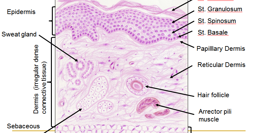
Thin Skin Histology Diagram
Skin (integumentary system) Histology skin thin diagram slides resolution high Illustrations: thin skin
Skin (Integumentary System)
Histology skin thin Skin histology nus pathweb annotations expand edu Histology dermis tissue epithelial sebaceous glands physiology corpuscles krause zapisano
Skin histology thin normal hematoxylin thick integumentary system drawings slides eosin trichrome
Skin histology leeds diagram layers sweat glands three dermis hypodermis muscle epidermis hair ac smooth pili arrector sebaceous functions foundSkin histopathology simple made introduction dermatopathology neoplastic dermpath dr Histology dermis tissue epithelial physiology sebaceous appendagesHistology (skin).
Skin (integumentary system)Dermpath made simple Skin: the histology guideSkin – normal histology – nus pathweb :: nus pathweb.

Histology of skin
Skin thin thick histology microscope drawings between integumentary system light differences specimensSkin (integumentary system) Skin histology integumentary system thin drawingsHistology slides database: january 2014.
Histology integumentary anatomy stain tissue types layers mallory trichrome cutisHistology (skin) .


Histology (Skin) - Part 1
Histology Of Skin | Faculty of Medicine

Histology (Skin) - Part 1

Skin (Integumentary System)

Skin (Integumentary System)

Illustrations: Thin Skin - General Histology

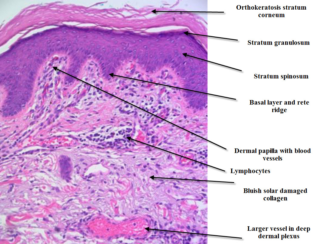
Dermpath Made Simple - Neoplastic: Introduction to skin histopathology

Histology Slides Database: January 2014

Skin (Integumentary System)